Renal Ultrasound
Basic principles of Ultrasound Examination
High frequency ultrasonic sound waves are sent through a transducer to the organs from a site where the transducer is placed on skin. The sound waves bounce off tissues like echoes and return to the transducer. The transducer coverts them into an electronic picture.
Different types of tissue affect the speed at which sound waves travel. Resistance to sound transmission varies with the nature of tissue. Fluid transmits sound with no resistance. No echoes are returned. That is why fluid is anechoic and appears dark. The sound is transmitted freely beyond fluid, posterior enhancement.
Pus is complex fluid and will produce low level echoes due to high protein content and tissue debris.
Sound does not travel well through air and that is the reason US examination is not optimal in abdominal examination. It is suitable for pelvic structure evaluation, where bowel can be displaced by filling bladder with urine. This is achieved by asking the patient to drink a large amount of water before the procedure. US is directed through fluid filled bladder to evaluate pelvic structures. Transvaginal US is also a method that allows visualization of pelvic structures without bowel interference and no need for filling bladder.
Bone does not transmit sound waves. Thus, US it is not suitable for evaluation of adult head. It is useful in children before the fontanelles are closed.
Calcified tissues do not transmit sound waves and are highly echogenic and appear white. Since they do not transmit sound there will be shadows posterior to the lesion (acoustic shadows).
Fat and hair are echogenic and appear white but are not associated with acoustic shadows.
Value of US in renal failure cases
Image atlas with examples for each type of renal failure
US is a non-invasive procedure useful in distinguishing the etiology for renal failure. It is critical for ruling out obstruction. When procedures requiring contrast like IVP and CT cannot be done, US of kidneys is very helpful.
Blood flow to kidneys can also be evaluated with doppler US. Flowing blood appears an echoic and dark. Vessels can be recognized in that fashion. With doppler, sound waves become audible based on the velocity of blood flow.
Right kidney can be evaluated through the liver. Left kidney is imaged in decubitus position. If unsuccessful, it is examined from back. If the spleen is enlarged it can be used as a window. These options avoid interference from bowel gas.
 |
Normal Kidney
- Normal kidney measures between 10.5 to 12.5 cms.
- Cortex is about 2.5 cms and has the same kind of echoes as liver.
- Central echoes are from fat surrounding renal pelvis and are white.
- Medulla is hypoechoic and appears dark. You can distinguish it from fluid filled calyces, as there will be no increased transmission of sound beyond.
- Renal pelvis is filled with urine and is echo free with posterior enhancement behind renal pelvis (next normal kidney).
|
 |
Normal Kidney
Kidneys appear normal in acute renal failure (hypovolemia or acute tubular necrosis). |
 |
Chronic renal failure (Medical Kidney)
- Kidneys are smaller than normal.
- Surface may be irregular.
- Cortex is thinned and is hyperechoic than liver due to scar tissue.
- Medulla is hypoechoic and appears dark. You can distinguish them from fluid filled calyces as there will be no increased transmission of sound as in fluid.
- Renal pelvis is filled with urine and is echo free. Note the posterior enhancement behind renal pelvis.
Kidneys are small, irregular and hyperechoic in chronic renal failure due to medical renal disease. |
 |
Hydronephrosis
- Dilated anechoic calyces with increased posterior transmission of sound.
- Hyperechoic fat delineating dilated calyces.
- Cortex is normal with similar echo pattern of liver.
Kidneys in obstructive renal disease show hydronephrosis. |
 |
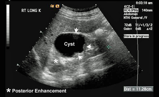
Simple Renal Cyst
Simple renal cysts are common. We call them simple when they are in the cortex, thin walled, the inner lining is smooth. The fluid is anechoic with no internal echoes and has good sound transmission with posterior enhancement. These cysts do not require further work-up. |
 |
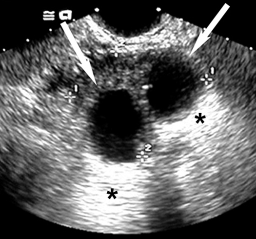
Complex Renal Cyst
Cysts where the inner lining is irregular, with septations and fluid has echoes are considered complex cysts. Complex cysts require additional further work-up to rule out cavitating cancer or renal abscess.
|